Learning How To Improve
Improve Efficiency With An Intraoral Camera In Every Room
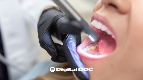
We play an essential part in improving oral health by educating our patients. Visual aids are required to make information accessible to the patient. Using an intraoral camera will help the dentist explain, define, and coordinate specific ideas that create urgency for treatment. Improve Efficiency With An Intraoral Camera In Every Room and see why dental offices everywhere are loving these.
Putting Intraoral Cameras to Use In Your Dentist Office
Intraoral cameras work best when each room has its camera, eliminating the need for the physician to seek it. Instead, the physician will be able to expand the image and discuss the status of the tooth by using a computer or an extra display.
For example, if a patient is dubious that an amalgam restoration has to be replaced, show the patient the stress lines, wear, and discoloration while comparing the situation to a healthy repair. In addition, taking images of the present status of the teeth can help the patient better understand their dental health. Finally, intraoral pictures are an effective instructional tool that physicians may utilize when discussing treatment choices with patients.
What Do Intraoral Cameras Do?
When taking shots, place the camera softly on the adjacent tooth. This will make the camera more steady, resulting in a better-focused image. In addition, make a technique for determining which quadrant will be photographed initially. Having a system to capture information will help the physician be more successful and efficient when numbering the teeth in the picture, leaving more time for teaching the patient. To get the most of the camera, take a shot of each tooth and surface, especially when creating a baseline for comparing future images. In addition, photos can show how the problem has progressed.
Begin the dialogue by emphasizing prevention, and then develop a dynamic teaching experience to encourage patient compliance. Once the photos have been obtained, identify the teeth in the shot for simple examination at a later date. At least once a year, provide updates with apparent changes in the tooth or tissues. The first or baseline pictures can be compared as the tooth changes to educate the patient on why treatment is required. Documenting the patients’ unique hazards, such as leaky restorative margins, stress fractures, and irritated gingiva raises the required therapy’s urgency.
Additionally, images should be taken during, before, and after therapy to allow the patient to witness the treatment procedures. Intraoral photographs are a strong tool for educating patients and can be utilized when talking with insurance companies.
Photos Boost Compliance
Using the camera to show the patient their bleeding pocket, irritated gingiva, coated tongue, or any other indicators of unhealthy tissue might be utilized to inspire change in the patient’s home care regimen. Our friends at High Desert in Grand Junction Co believe that educating your patient with a snapshot of their oral cavity will empower them to take better responsibility for their oral health. Describing the patient’s existing tissues compared to healthy tissue will assist in developing healthy oral hygiene practices. Images can also be used to congratulate and encourage the patient for their achievements by following the provider’s homecare advice.
Contact Us Today
Using an intraoral camera improves the patient’s knowledge of the tooth’s diagnosis. Because our objective as dental professionals is to assist our patients in attaining oral health, boosting chairside acceptability is critical to keeping our patients healthy. Implement an intraoral camera into your everyday patient education to take your chairside teaching to the next level. Call Digital Doc today and get you and your dentist’s office a new intraoral camera.

Blog body text

