The Key to Modern Day Root Canals

essentials to modern day root canals

The Key to Modern Day Root Canals

The modern dental patient expects their dental care provider to use the latest technology, deliver better outcomes, and be willing to schedule procedures at the convenience of the patient. Dental care providers are scrambling to improve the way they deliver their services in order to meet these expectations of their patients. This article discusses five essentials that can help to deliver modern-day root canals that meet all the expectations of patients.

XTG Handheld X-Ray

If you’ve used Digital Doc’s XTG Handheld X-ray, you’ll wonder how you managed to perform root canals in the past. This Xray2go allows you to remain in the operatory and take radiographic images of your patient while they are in the dental chair. Root canals are one of the procedures that patients don’t look forward to undergoing, but with this tool, you can increase the convenience factor for your patients because they don’t have to be shuffled to the radiology room and then back to complete their treatment.

The handheld x-ray also gives you high-quality images that can help you to deliver treatment outcomes that can only be dreamed about by those using the traditional x-ray machines.

An IRIS Intraoral Camera

While endodontists largely treat patients who have been referred by dentists, the records sent along with the referral may not provide an accurate depiction of the patient’s tooth before the root canal treatment is undertaken.

It is therefore helpful for you to have an intraoral camera like the IRIS USB 2.0 Chair Dental Camera so that you can take high-quality images to complement what has been captured by the XTG Handheld X-Ray.

Effective Communication

It is one thing to communicate, and it is another thing to communicate effectively. For example, you wouldn’t use the same diction that you use when communicating with a referring dentist to talk to a patient, would you?

It is important for you to communicate effectively with all parties, such as your patients, the specialists and referring dentists if you want to deliver better root canals.

Luckily, modern digital dentistry equipment like the LUM and XTG Handheld X-Ray make it possible for all parties to be on the same page. For instance, you can take images with your IRIS Intraoral camera and show the patient what exactly you are going to fix during the root canal treatment. The same images can be used if you need to communicate with referring dentists so that everyone is kept in the loop during each stage of the treatment.

Knowledgeable Staff

Another important factor for endodontists who wish to perform modern-day root canals that meet all the expectations of patients is knowledgeable staff. Your staff should be conversant with the workings of all your modern equipment, such as the Xray2go. They should also be familiar with how that equipment delivers outstanding outcomes for your patients.

Such mastery can only happen if you, the endodontist, set the example and arrange to have your staff trained each time you introduce new technology or methods of delivering patient care. Your staff will then convey their expertise to the patients, and the net result will be superior root canal outcomes when compared to other offices.

Once you have all the essential components above in place, there will be nothing standing in the way of delivering modern root canal treatments. As you may know, word of mouth spreads fast, so your practice will grow as a result of all the positive reviews made by any patient who undergoes root canal therapy at your office.

The Safety of Portable Dental X-ray Machines

the safety of portable dental x-ray machines

The Safety of Portable Dental X-ray Machines

Dental X-ray devices are extensively used in dental treatment just like the LUM Sub-Enamel Illumination device. In order to serve patients well, clinicians should be aware of and prepared for the hazards of using x-rays. X-rays emit radiation, which can prove to be significantly risky for patients if not used appropriately. Operating an x-ray machine is not as simple as pushing a couple of buttons, technicians should train themselves properly to use x-ray devices. The same is true for portable handheld x-ray devices.

Digital Doc XTG is an excellent portable x-ray system. It is a high frequency, advanced dental x-ray device. With a 60kVDC and 2mA tube current, it is built to produce high-quality x-ray, for both film and digital sensors. The devices can also be mounted on an optional tripod or on arms. Aside from improving efficiency in patient diagnosis, it is safe and easy to use the device for clinicians. The rest of the article will outline the key guidelines and safety precautions to follow while using the device.

Check the X-ray Device Package

First things first; as soon as you get the device open and assess all the components provided in the packaging for damage. The standard device packaging will consist of the device, power cable, the long cone, user manual, backscatter shied, the remote control switch (optional), and battery charger.

You must carry out the following preliminary checks:

  • Check if the device has serialized device label (usually on the back)
  • Inspect Collimator Cone and Backscatter Shield for any shipping damage
  • Check if Exposure Switch is functioning freely
  • Ensure that Device Housing has no cracks or fractures

You should also keep the following important points in mind:

  • Do not open the housing (enclosure)
  • Warranty will be rendered useless if you disassemble the main apparatus
  • Only trained personnel should repair the device
  • Gather information from an authorized distributor only

Dental X-Ray Radiation Prevention

Not only should you register properly and comply with all applicable regulations, but you should also consult the relevant state, provincial, and local regulations which govern radiation protection and use of x-ray equipment. The operator of the device should carefully follow all enforced regulations in relation to pregnant or expecting to be pregnant women. Operators of the device should also be completely familiar with the industry safety recommendations and permissible doses. To ensure optimal radiation backscatter protection you should:

  • Position the backscatter shield at a correct angle to the operator
  • Keep backscatter shield close to the patient
  • The patient tilts their head when required to do so
  • The operator stays immediately behind the permanently attached backscatter shield, in the Significant Zone of Occupancy

Only begin using the XTG when you are sure there will be no interruptions, putting other people under risk of being exposed to radiation. The assistant should also wear proper protective gear. In using Position Indicating Devices (PIDs), models allowing the operator to position the unit at the correct angle should be preferred. The depressed trigger can be used to end exposure at any given point. Low-class image detectors should not be used.

Duty Cycle and Usage of Dental X-ray

Xray2go has a maximum duty cycle rating of 1:60, and is designed in a manner to avoid overheating from excessive use. However, the device should not be used in an area with flammable material. The optimal battery voltage level for using XTG is 22.2 VDC, and it should not be operated on low voltage levels.

Proper Cleaning of Handheld X-ray

Before cleaning, you should ensure that the battery charger is unplugged. Keep the power of the device off while you clean it. To clean the device you can use non-alcohol based disinfectant wipes, a cloth with disinfectant liquid, or spray. The device is not water-proof, so care must be taken to not dampen it with any form of liquid. XTG should also not be sterilized.

Storing and Transporting the dental x-ray

For long term storage, the device should be stored in a place which is not affected by air pressure, temperature (cool), ventilation, humidity (dry), salt, dust, sunlight, etc. Be sure to avoid dropping or hitting the device while transporting or storing it.

Regular Maintenance on XTG Handheld X-ray

A technician should make an annual maintenance check on the device. You should also be wary of the accessories you use; if non-recommended accessories are used, they may damage the device.

Understanding Caries Detection Devices

understanding caries detection devices - digital doc

Understanding Caries Detection Devices

Over time, medicine has moved away from aggressive surgery to minimally invasive procedures as well as placing more emphasis on prevention and early detection. This change has been triggered by the need to lower the rates at which complications develop after surgery, as well as the need to reduce recovery time and the pain experienced by patients. Dentistry has also taken the same trajectory and caries detection devices, such as the LUM, have been introduced on the market in order to make it easier to catch caries early and reverse this condition before it costs patients their teeth. This article discusses some of the shortcomings of the traditional caries detection technology and also explores the benefits that the new crop of caries detection devices bring to the field of dentistry.

The Shortcomings of Traditional Caries Detection Technology

Radiographs: Radiography has been used for long in the fight against caries. However, this technology has one big weakness. Radiographs tend to be effective in alerting a dentist about the presence of caries once those caries have advanced and reached the dentinoenamel junction. In other words, radiographs can only detect caries with a high degree of accuracy once the bacteria causing caries has done so much damage that the dentine is starting to be exposed. This is unacceptable for dentists interested in minimally invasive dentistry since such severe damage requires invasive remedies or interventions.

Dental Explorers: dental explorers have also been used for long to examine the surface of teeth in order to find out which portions have caries. However, dental experts have been discouraging the use of these devices because the sharp tip of the explorer can compromise the portions of the teeth that are re-mineralizing. This can inadvertently give caries a chance to advance further.

The Benefits of Caries Detection Devices

The age of digital dentistry has ushered the dawn of caries detection devices like the LUM. These devices offer a variety of advantages or uses, such as the following;

Checking for caries before sealants are placed: Caries detection devices provide sub-enamel illumination so that dentists can use their findings to make various decisions. One of these pieces of information collected with the help of LUM is the state of any caries on the teeth before dental sealants are placed. This information can be captured more effectively once the caries detection tool is used alongside a high-quality intraoral camera, such as the IRIS HD USB 3.0 Dental Camera. The images captured can be analyzed thoroughly so that any caries detected can be treated before the sealants are put in place.

Assessing Re-mineralization Measures: Caries detection devices can also be used to monitor whether the measures to re-mineralize the teeth of a patient are working as expected, or not. The information gathered can be used to change the treatment plan if tangible results aren’t forthcoming, or a specific approach can be retained if it is bringing the desired outcomes. In this way, the time that would have been lost waiting for an ineffective method to bear fruit can be saved.

Monitoring Sealants: Caries detection devices have also shown their usefulness as a tool to help dentists to monitor the edges of dental sealants as time goes by. Any deterioration can be tracked so that a timely decision to replace the sealants can be made. This proactive approach could not have been possible if caries detection devices didn’t exist.

As you can see, caries detection devices like the LUM sub-enamel illumination device can add a whole new dimension to the ease with which your dental office can catch caries early. However, you should use these devices together with the other caries detection systems for better results. Contact Digital DOC for a virtual or in-office demo so that you can grasp how easy your work will become once this tool is available to you and your staff.

How to Improve Case Acceptance in the Modern Dental Practice

How to improve case acceptance in modern dental practice

How to Improve Case Acceptance in the Modern Dental Practice

Your office may have experienced, and skilled staff, the latest dental technology, but that may come to nothing if your case acceptance rates are dismal. Read on and discover how to improve case acceptance in the modern dental practice.

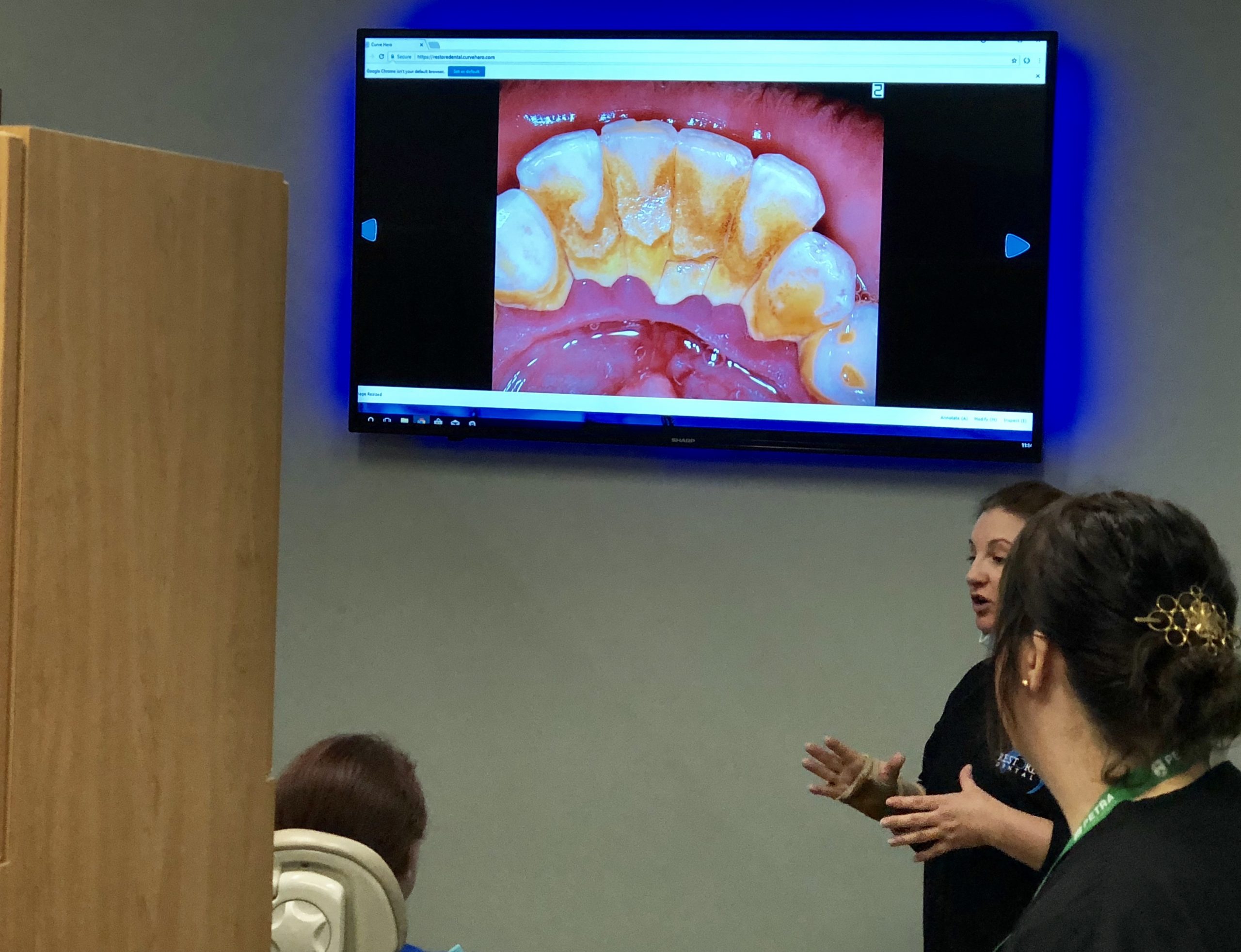
Use Pictures, Rather Than Words

You can go on and on about treatments, but a patient may still say no to a necessary or recommended dental treatment procedure if they don’t see the importance of it. Why not just tell your patient, but show them what you can do for them.

If you use the appropriate images to show your patient a firsthand view of what the problem is, they’re likely to change their mind. For example, the patient may require deep cleaning instead of the routine cleaning that he or she had come for. Use the IRIS HD USB 3.0 Dental Camera to capture images of the affected teeth. Beam those images onto a large screen within the operatory so that the patient can see how serious the situation is. Your patient will then realize that your recommendation is indeed necessary, and he or she will likely move forward with the recommended procedure.

Emphasize Same Day Treatment

We live in a very busy world, and everyone seems to be pressed for time. How can your dental practice be more considerate of your patients’ time?

Case acceptance in the modern dental practice can increase significantly if same-day services are emphasized. Recent technologies, such as the XTG Handheld X-Ray from Digital Doc, have collapsed the time that was initially required for patient care. The handheld X-ray allows the dentist to get instant x-ray images that can be displayed on a screen and reviewed right then and there. So, any needed dental work can be completed during one visit instead of having the patient come back for additional appointments.

In short, digitize your dental practice so that same-day services become the norm. You will see an increase in case acceptance rates at your practice.

Show Your Dental Patient Urgency

Another way to increase case acceptance in the modern dental practice is to stress the risks of delayed treatment to your patient. For example, a patient who comes into the office with a toothache should be told that urgent treatment is needed before more damage is done, which will cause additional dental procedures like a root canal.

Fortunately, sub-enamel illumination technology like LUM can be paired with an IRIS Intraoral Camera to show the patient how extensive their dental problem is. Consequently, it is likely that the patient will agree to immediate treatment once they fully understand the urgency of the situation.  

Adress Your Patient’s Fears of the Dental Treatment

Sometimes, case acceptance rates can take a tumble when you don’t address your patient’s fears. For example, a patient may postpone a needed surgical procedure simply because he or she fears that the procedure will be so complex that there will be complications and the recovery time will be longer than expected.

Your staff can learn to integrate language that removes fear from your patients during consultations. For example, as you discuss the images taken using the IRIS LTE USB 2.0 Dental Camera beamed on a screen, inform your patient that their tooth decay treatment is a routine procedure and they’ll be back to normal in no time. Additionally, you can explain that you have done it countless times without any problems. Addressing your patient’s fears will help increase the case acceptance rates of your dental practice.

Refrain from the Money Talk

Your work as a dentist is to educate and treat patients. Focus on doing just that and avoid discussing the cost of the procedure. Let the front office or billing department discuss this with the patient. The best thing you can do is educate your patient about their necessary treatment. Make the best use of the IRIS Intraoral Cameras to help the patient understand why the urgent treatment is needed.

As you can see from the discussion above, case acceptance in the modern dental practice largely depends on how you present the case to the patient and their understanding of the necessary treatment. Digital Doc can help you to digitize your practice so that better patient care can be offered efficiently without sacrificing attention to detail. Contact us today for a demonstration of how our products can help to skyrocket your case acceptance rates.

Traditional Dental Impressions vs. Digital Dental Impressions

Digital Doc - Traditional Dental Impressions vs. Digital Dental Impressions

Traditional Dental Impressions vs. Digital Dental Impressions

If you would like to provide dental restorations services, such as dental crowns, in your office then you’ll need to take dental impressions first. You can offer your patients traditional dental impressions or you can use digital dental impressions. This article discusses the two types of dental impressions you can provide in your office.

Traditional Dental Impressions

Traditional dental impressions are obtained by placing dental putty in the mouth of your patient. This putty hardens slowly. Your patient will bite down on that so dental impressions are left in the substance. 

Thereafter, a lengthy process of preparing the mold for the restorations begins and sometimes, retaking the dental impressions may be required to fix any errors made during the previous session.

There is a nearly universal dislike of this traditional dental impression capture method by both dental care professionals and patients alike because the putty triggers the gag reflex in patients and makes for unpleasant dental visits.

Additionally, traditional dental impressions are time-consuming for the patient and for dentists. In addition, you may have to repeat all these steps if the restoration does not form correctly for your patient. 

All these issues prompted a better way to get dental impressions, and digital dental impressions were brought to the market by Digital Doc and a few other manufacturers.

Digital Dental Impressions

Digital dental impressions are taken by the use of an intraoral scanner like the Heron IOS Intraoral Scanner. This scanner captures images of all the intricacies of your dentition and creates 3D images of your dental structures so that a restoration can be fabricated. Digital dental impressions have a number of advantages over the traditional way of getting dental impressions.

First, less time is required to take the impressions simply because dentists can move the Heron Intraoral Scanner within the mouth and in a minute or so the required impressions are secured. This is unlike the traditional dental impressions that require you to wait for a long time before the putty sets so your patient can bite into it.

Secondly, your patient will have a more enjoyable dental visit since they will not have to spend a lot of time with unpleasant-tasting substances in their mouth. Digital impressions eliminate all that and the process is completed quickly, as already mentioned earlier.

Additionally, the digital dental impressions obtained are accurate and hardly ever require a retake since the images captured are sent directly to the fabricator so that the restorations made conform to the structures in your mouth. The chance of human error during the capture of the images or the fabrication of the restorations is reduced since everything is done digitally.

Furthermore, you will be pleased to learn that digital dental impressions are eco-friendly if you are concerned about the environmental footprint of everything that affects you. Digital dental impression technology saves the energy that would have been used to store the materials used to get the impressions. Numerous materials, such as dental impression trays, are also not needed when taking digital dental impressions.

As you can see from the discussion above, digital dental impressions have so many advantages over traditional dental impressions. If you’d like to acquire Digital Doc products like the Heron IOS Intraoral Scanner in your office and provide excellent services to your patients, contact Digital Doc today for a consultation!


Best Intraoral Camera 2018

Lum Intraoral Camera

IRIS Intraoral Camera Wins 2018 Product Award

Dental Advisor submitted the IRIS Intraoral Camera to nine dentistry consultants and asked them to subject the camera to a clinical evaluation over a period of four months. Their verdict? IRIS was given a 96 percent clinical rating overall by that team of consultants. Read on and learn about this camera and what that 2018 evaluation entailed.

Overview of the IRIS Intraoral Dental Camera

The IRIS intraoral camera is a user-friendly dental camera which has a five-point focus wheel. A dentist can use this focus wheel to determine how large or how small the area captured in the image should be. Eight positions are available for the user to hone in on the type of image to be captured, such as clear images of the arch, entire smile, the full face and so many other areas of interest.

The IRIS intraoral camera comes with a cable which is 15-feet long so that the user isn’t constrained when moving about to capture a variety of images when a patient is in the operatory.

The camera comes with two capture buttons to suit operators who are either left-handed or right-handed. This has the added benefit of easing the capture of images even when the camera is being turned around in the mouth of the patient.

Sony’s hi-resolution CCD and eight-point LED lighting makes the camera capture clear and sharp images with true colors.

The IRIS intraoral camera requires 1GB RAM, a 2.0 USB interface and at least 2.0 GHz as its computer system requirements. IRIS now makes a version for MAC computers as well. The camera is charged via the USB port on any PC.

Other Reasons why more Dentist’s Choose IRIS 

HD intraoral camera

  • High Definition 720p resolution
  • Motion activated auto On/Off
  • 8-point true white LED array
  • One-piece slim design soft tip
  • Dual capture buttons for left or right hand use
  • One-touch 5-position electronic focus for a macro to infinite viewing with OSD
  • High performance impact and chemical resistant seamless body
  • USB 3.0 connector that fits standard delivery units


What the Consultants Said

The Stainless Steel Wand. The consultants agreed that the sleek, stainless steel wand is ideal for a clinical setting since this material is easy to maintain at the high standards of hygiene expected in a hospital setting. The stainless steel can also stand up to the harshness of some of the cleaning products commonly used in dental offices and hospitals. This is unlike plastic wands that soon wear due to the impact of the cleaning products typically used in a dental office or hospital setting.

Camera Software. IRIS also received kudos because the software upon which it operates was found to be easy to integrate into most of the commonly used patient management systems in dental offices.

The Lens. The 90° lens makes positioning the camera to capture images easy and intuitive. The images captured will, therefore, be sharp and clear due to the natural orientation of the lens.

The Barrier Sheath. Not many cameras can work satisfactorily when a barrier sheath is in place. Some models don’t even allow a barrier sheath to be installed. The IRIS intraoral camera is one of those few dental cameras whose image quality isn’t affected even with a barrier sheath in place.

The Lighting. The LED lights were found to be more than adequate to illuminate the field as images are taken. Those lights could also be turned off as a user is capturing images that have been backlit by x-rays.

The Consultants’ Conclusions

11% of the consultants who evaluated the IRIS Intraoral Camera said that it was similar to other intraoral cameras available. However, the majority of the consultants were of the view that the IRIS intraoral camera was superior to the other products on the market. All the consultants were in agreement that they would recommend the IRIS Intraoral Camera to any dental practice which needed equipment. They also recommended that dental practices using the IRIS camera should install a cable kit in all the operatories where they expect to use the IRIS intraoral camera so that moving the camera from one operatory to another is seamless. All in all, Dental Advisor relied on the report of the consultants to award IRIS Intraoral Camera the 2018 Product Award.

Help Patients Say Yes to Treatment

Help  Patients Say Yes to Treatment

A recent statistic states that there is $32 billion in treatment approved for financing Care Credit still waiting to be scheduled – why?

Why are patients not ready to purchase? Are my services not good enough? Are they going to find another dentist?

Sometimes we forget our patients are consumers too. The reason why they may not be ready to purchase is most likely because of the thought process behind consumer purchases.

Think about the last time you purchased something. Maybe you bought a flat screen TV or a brand-new car or limited addition shoes you.

What made you buy it? Was it the product itself? Was it the look, the feel or the design of the product? Was the product on sale? Did you need your purchase or did your emotions play a role in your decision?

The average consumer today buys based on their emotion. So how do I get my patients to buy from me when there seems like there’s little room for improvement?

Well, many doctors will say “My intraoral camera image is good enough.”  But good enough for who?  The highly educated consumer sitting in your chair with a $1,150 cell phone in their pocket? 

The average consumer today expects the best experience possible, so they will notice if your office equipment is outdated. New technology, like intraoral cameras, is constantly improving so there’s always room for improvement, Consider upgrading your cameras and monitors in your office to make their experience even better.


How an Intraoral Camera Can Have a Huge Impact on Your ROI

How an Intraoral Camera Can Have a Huge Impact on Your ROI

So you tell your patient he needs to get a couple of crowns or fillings after conducting a routine cleaning and exam.

Your patient looks at you uncomfortably and says, “Maybe I can decide on my next appointment, Doc. I don’t have any pain, at all.”

Then you see the distrust in his eyes and tell him reassuringly, “Sure, no worries. I just hope the problem with your teeth would not have grown worse by then.”

He then reaches for his wallet, murmurs “Thank you,” and rushes off.

Sound familiar?

Now, you’re not sure if he’s ever really coming back, even after six months when he’s supposed to have his next routine cleaning appointment.

But have you ever considered getting an intraoral camera for cavity detection and other crucial aspects of your practice? If not, consider the following advantages:

  • Early detection of dental problems – The smallest signs of a growing dental issue such as hairline fractures can be easily detected with an intraoral camera.
  • Accurate diagnosis – With the crystal-clear, sharp images that you can take with an intraoral camera, you’ll be able to make precise diagnoses and recommended the right treatment every time. For more difficult cases, you can easily confer with other experts since high-resolution images will be available during consultation.
  • Patient education – You can virtually do “show and tell” in real time. This means you can take your patient on a “dental tour” of his or her mouth, showing areas where gum disease may be a concern, as well as plaque and other early signs of tooth decay.
  • Improved doctor-patient relations – With an intraoral camera, your patient knows right away that you’re not “making stuff up” in terms of his or her oral health. Images of patients’ teeth and gums taken in real-time means they see exactly what you see, and you can easily point out areas they need to be concerned about.
  • Increased case and treatment acceptance – Using images taken with an intraoral camera means your patients won’t need a lot of convincing to make decisions for their treatment because they know and have seen the state of their dental health.
  • Filing or future case documentation – The availability of images from each patient case means you need not spend a lot of time writing up descriptions of each case. You’ll also save on office space as you can avail of both traditional PC-based and cloud-based information storage options.
  • Stronger dental insurance claims – When there are problems with dental insurance claims, there really is nothing like crystal clear pictures painfully detailing your patient’s dental problem and the need for treatment to reassure insurance companies that the claim being made is well-founded and legitimate.
  • Enhanced practice reputation – Having an intraoral camera around can have an immense impact on your credibility and integrity as a dental practitioner. Your patients trust you, and you can also trust them to refer you to friends and family.

The key takeaways here, in terms of the profitability of your practice are, of course, improved relations with your patients and case acceptance.

So now that you are familiar with how an intraoral camera can have a huge impact on your ROI, it’s high time you do some more research and shop around. How many do you need? What can I expect in terms of a % of growth in Year 1?

You can start off by exploring the IRIS intraoral camera series, including the IRIS USB 2.0 Chair or IRIS LTE USB 2.0 intraoral cameras which both offer flexibility and ease of use as well as sharp, high-resolution imaging results. Contact us today to schedule a free demo.


Selecting the Best Caries Detection Tools for Your Dental Practice

Selecting the Best Caries Detection Tools for Your Dental Practice

There’s no question that caries or cavity detection is one of the most important tasks of dental practitioners. In fact, a lot of patients will only go to a dentist if they feel pain – the proverbial toothache – or if they see physical signs in their teeth such as bleeding and/or discoloration.

This is primarily the reason why it is so important for dentists to ensure they have the education, training, skills, experience and current technology required to establish a sound and respected clinical practice.

Therefore, in light of this common need among patients, selecting the best caries detection tools for your practice is paramount to ensure you get accurate information in order to arrive at a correct diagnosis and treatment recommendation.

Below is a list of detection tools used in dental practice:

1. Detection Dye

The caries detection dye or disclosing agent has been an important supplemental ingredient in the use of the dental explorer, otherwise known as the “sickle probe,” and spoon excavator.

Traditionally used to prod and probe teeth for the presence of cavities, the dental explorer requires the use of caries detection dye as the latter is usually effective in detecting caries after teeth have been subjected to a thorough cleaning.

Detection dye can accurately differentiate between non-remineralizable and remineralizable dentin and is considered one of the more cost-effective caries detection tools.

As you may know, staining solution is applied to the thoroughly dried and isolated area and usually left for 10 seconds and then rinsed off (sometimes immediately, according to the manufacturer’s recommendation).

Areas that turn scarlet red (or even dark blue or green, as the case may be, depending on the formulation of the solution) are identified as carious dentin and are subsequently removed with a round bur and slow speed handpiece and/or a spoon excavator.

The problem with this method is that it can lead to false positives because circumpulpal dentin and that which is found in the dentino-enamel junction are less mineralized.

2. Laser fluorescence (LF)

Used to measure the extent and depth of decay, laser fluorescence technology can be used both for diagnosis and treatment.

It is known that bacteria fluoresce when subjected to various wavelengths of light, a premise upon which LF technology is built upon. When carious structures are detected, the device gives off an audio signal indicating the extent of caries.

Current improvements in the technology offer greater accuracy in detection, although the American Dental Association (ADA) recommends that LF is used in conjunction with other caries detection methods for better and more accurate diagnosis and treatment.

The effectiveness of this tool is highly dependent on the level of expertise/training of the dental practitioner.

3. Intraoral cameras

More efficiently used together with dental imaging technology and/or dental management software, intraoral cameras offer precise, real-time images to detect dental problems, including caries.

Connected to a PC monitor or TV screen visible to the patient, intraoral cameras produce high-resolution images and enable dentists to see hard-to-reach sections of the oral cavity. Depending on the model, they are generally lightweight, flexible and easy to manipulate.

The LūM Transillumination, by, Digital Doc, attaches to the IRIS intraoral cameras not only taking external photos of the teeth, gums and general mouth area but also features sub-enamel illumination showing nearly X-ray like images. This technology is extremely powerful when presenting a treatment plan to your patients.

Choose your technology

While there are other tools available for the early detection of dental caries, both old and new, it is up to you, the dental practitioner, to decide on which will work best for you.

In making your decision, always take into account the level of accuracy the tool you use produces. It will directly impact the diagnoses you will be making, and the treatments you will be subjecting your patients to. Remember, quality over quantity goes a long way in dentistry. Choosing the right equipment will earn you trust, and raving fans in your patients.

In short, the success and reputation of your practice depend on it, so be sure to do your homework.  

SEE More. DO More.

Dental Instrument Quality Test Guidelines

All of the cliche sayings like, “time is money”, and “you get what you pay for” come to mind when we talk about dental instrument quality test guidelines. In this short video with our President, Brett Wilson, we share why quality test guidelines are important for your patients safety. Let us know what questions you have! 

H1 blog title

H2 Different from title but including focus keywords

Blog body text 

Blog body text