Why Choose a Dental Sensor

5 Benefits Of A Dental Sensors

Why Get A Dental Sensor

Why Choose to Purchase a Dental Sensor For Your Practice

Dental x-rays are an important component of normal dental checkups. Not only should the surface portions of teeth be examined, but so should the inside areas as well. There is no doubting that traditional dental x-rays have aided a large number of individuals. However, technological improvements have enabled engineers to design gadgets that deliver immediate, HD digital imaging. Going digital can elevate your practice, but why should you get a dental sensor?
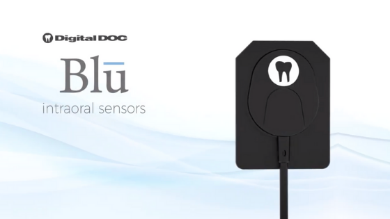
The Blū

To start, our advanced dental technology team shares the advantages of our newest sensor, the Blū. Our revolutionary intraoral x-ray sensor will take your practice to the next level. Blū is long-lasting, comfy, and, most of all, inexpensive. The Blū intraoral sensor, with its crystal clear pictures, will serve as your second pair of eyes during the treatment planning phase. So, if you haven’t already gone digital, start with Blū. It certainly is everything you could want in a digital dental sensor and more. 

Dental Sensors Provide Great Visuals

Dental technology helps doctors and patients communicate more effectively. Dr. Green, Parker CO dentist, explains that this allows dentists to show their patients the problem rather than telling them about ir. It’s difficult to agree to costly procedures when the problem isn’t seen in low-quality images. The high-definition imagery of Digital Doc’s Intraoral Dental Sensor Blū will help you make a diagnosis and justify your treatment approach.

Dental Sensors Improve Efficiency

All of us in the dental field know that a standard exam’s x-ray section might be the most time-consuming element of the procedure. Luckily, dental sensors can help with that. They have minimal setup and modifications. Thus, this reduction in setup time improves overall office productivity and allows you to focus on what really matters: the patient.

Get Digital Doc Support

It is our responsibility as an industry leader to supply you with high-quality products and superior service. With the assistance of Digital Doc Support, you will have no trouble implementing your new dental sensor. Our virtual training will teach everyone in your workplace how to reap these benefits. You can always rely on us if you have any queries or issues concerning your Digital Doc products. Contact us today to learn more. 

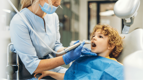
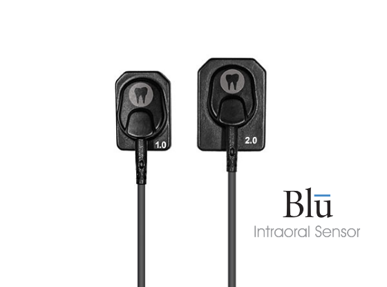
Increase Comfortability For Both Patients and Team with Dental Sensors

It’s understandable that traditional x-ray methods can be uncomfortable for patients. Thus, Digital Doc’s Blū Intraoral Sensor comes in the perfect size, making for the best patient experience possible. You can choose from a 1.0 and 2.0. Each sensor is about 4.8 mm thick, with the dimensions of 31.3 mm X 42.9 mm. 

Contact Digital Doc To Learn More

If you’re interested in our Digital Doc products, please reach out to our team. We can schedule a virtual demonstration today. Additionally, we can discuss any of our products – dental cameras, X-ray devices, accessories, and more! Follow us on InstagramFacebook, and YouTube to learn more!

Blū Digital X-ray Training

blu digital x-ray training - Digital Doc

Blū Digital X-ray Training

Blū Digital X-ray Training for Your Practice

Welcome to the training of Digital Doc’s Blū Digital X-ray sensor. Today, we will cover the barrier’s placement, use of the x-ray positioning system, proper placement of each type of x-ray, proper disinfection, storage, and technical support included with your sensor.

The Digital Doc Blū X-ray Sensor delivers low noise, high contrast images for fantastic diagnostic reliability. The x-rays captured on our sensors are transmitted instantly to your imaging software, where they are ready for your diagnosis. 

Different Sensor Sizes

Patients come in multiple sizes, and so do our sensors. Two sensor sizes give you the flexibility to get the right image for every treatment situation. 

Our size 2 sensors are standard size for adult patient bitewing x-rays and posterior periapical x-rays. Our size 1 sensor is ideal for pediatric patients, anterior PAs, and patients with a small mouth or large tori. 

Barrier Placement

To prevent cross-contamination of the sensor between patients, you always want to use a barrier or sheath on the sensor before putting it into the patient’s mouth. Using the recommended barrier for your sensor, slide the sensor into the barrier, pushing it all the way to the end. Make sure that the cable is covered as well. When removing the barrier sensor, you want to make sure you avoid pulling on the cord. 

Sensor Placement Per X-Ray

Now that our sensor is properly covered, we can place the sensor on our x=ray positioning device. For any radiograph, seeing the interproximal contact clearly is important for caries detection. Digital doc’s x=ray positioning system helps ensure you’re using the proper paralleling technique while acquiring your image. Place the sensor holder with the sensor’s flat side towards the ring and the corded side of the sensor toward the center of the mouth. You always want to aim the x-ray machine at the flat side of the sensor. Make sure the cord is directed out of the mouth, not towards the throat. You should be able to see the sensor directly through the ring. 

Our x-ray positioning system is set for the upper right PA. Place the sensor on the lingual of the upper molars. The sensor will tilt towards the sensor o the pallet. Have the patient bite down. Make sure the ring is as close to the cheek as possible. Place the x-ray cone up to the ring.

blu digital x-ray training - Digital Doc

Dental X-Ray Exposure

For lower posterior PA, retract the patient’s check for increased visibility into f the mouth. Place the sensor between the lingual side of the lower molars and tongue. Again have the patient bite down. Make sure the ring is as close to the face as possible.

For the upper anterior periapical x-ray, scoop the sensor into the mouth’s pallet and place it behind the upper anterior of the teeth. Have the patient bite down. If needed, they may use their hand to hold the bar steady. Push the ring as close to the face as possible.  For the upper anterior, the ring will be very close to the nose. Line up the x-ray tube with the ring and expose the x-ray.

For horizontal bitewing x-rays, place the sensor between the tongue and the teeth. Align the front end of the sensor towards the midline to follow the mouth’s curve and help with getting open contacts on your image. Have the patient bite down. Press the ring as close to the face as possible. Line up the x-ray with the ring and expose your x-ray.

Proper Disinfection of Sensor

After you’ve removed the barrier, use recommended disinfectant wipes to wipe the sensor and cable. Ensure you are wiping towards the sensor, not away from the sensor, to ensure you are not pulling on the cord. 

Storing the Sensor

A wall-mounted sensor holder is always recommended. Place the sensor in the holder and wrap the cord around so that the cord is not coiled tightly or kinked in any way. 

Warranty and Technical Support

Your x-ray sensor carries a 1-year manufacturer warranty. See our website at www.digi-doc.com for warranty details and details on our extended care plan.

For sensor technical support: please call the Digital Doc Corporate office located in Northern California at 1-800-518-1102 between the hours of 9:00 am and 5:00 pm CST for any technical assistance you may have. 

Subscribe to our Digital Doc YouTube channel for more videos on our Digital Doc products.

How Dental Technology Can Relax Patients

how dental technology can relax patients

How Dental Technology Can Relax Patients

How Dental Technology Can Relax Dental Patients

Fear of visiting the dental office is an issue people face all over the world. In a recent worldwide survey of about 18k people, more than 60% of respondents said they were suffering from dental anxiety and fear of the dentist. More often than not, patients are avoiding their dentists altogether. Fortunately, new advanced dental technology improves the overall patient experience. Below, we explain how dental technology can relax patients and increase case acceptance. 

Understanding Your Patients’ Needs

In the dental office, anxious patients usually show irritability, are not agreeable and are more likely to respond to pain than their compound counterparts. These responses are troublesome not only for the patient but also for dental professionals. In order to prevent unpleasant patient encounters and ensure quality treatment, dentists and their teams need to practice meaningful ways to assist patients with dental anxieties to help them resolve and conquer their fears. Here are a few ways you can incorporate these technologies into your practice.

how dental technology can relax patients

Teledentistry

Teledentistry has become increasingly popular during the COVID-19 pandemic. To reduce potential exposures while keeping oral care, many dentists are utilizing Teledentistry. To learn more about Teledentistry, read Facts About Teledentistry

Now that most Americans have cell phones, tablets and laptops are now fitted with high-precision cameras resulting in ultra-high-definition images; patients may obtain dental appointments from the comfort of their own homes without the stress and anxiety associated with them.

Benefits of Teledentistry

With Teledentistry, new patients will no longer have to waste time in the waiting room while you consult video instead. There is no need for dental assistants to donate and drop off personal protective equipment (PPE) for each new patient on board.

You may also have an extra degree of comfort and protection when it comes to periodic patient checks to monitor your home treatment or the healing of a newly implanted implant. There was no waiting room, no travel, and, again, no PPE required. Teledentistry helps reduce fear while ensuring their safety during a pandemic.

Virtual Reality

One of the most daunting aspects of dental anxiety that practitioners face today is the fear of the patient having to experience unnecessary discomfort during the operation. Despite advancements in medical methods and technology, the majority of people are still delaying or even fully refusing dental care due to the fear and anxiety surrounding the pain. 

Doctors everywhere are putting patients at ease with virtual reality (VR). Of course the distraction takes away from the pain. By encouraging a patient to concentrate his or her attention on other thoughts, less attention is available to the pain.

Virtual reality (VR) uses advanced technology to create virtual environments (VE) that allow patients to immerse themselves in an immersive, simulated world. These sophisticated devices communicate with the VE at several levels, stimulating sights, sounds, and motion to promote immersion in the virtual world to improve pain distraction.

Our friends at Park Hills Family Dentistry, a dental office in Lexington, KY, add that studies have shown that patient participation in VE decreased their recorded levels of pain during dental work.

Advanced Dental Technology

Thanks to advanced dental technology, doctors are able to provide patients with a more accurate diagnosis, quality treatment, and maximum comfort. As a dentist, you need technology you can rely on. The IRIS intraoral cameras easily capture high-resolution images. You can show patients and insurance companies exactly what you’re seeing. As a result, patients are more likely to say yes to your treatment plan. Contact us today to find out which IRIS intraoral is right for your practice. 

The XTG handheld x-ray device is another practice staple. Dentists everywhere enjoy our portable device that not only shoots pristine images but also has less radiation than traditional dental x-rays. The XTG comes with a shoulder strap and portable batteries. Taking dental x-rays has never been this easy!

Lastly, our new digital x-ray sensor, Blu, is your second set of eyes. Patients love Blu because it’s comfortable and noninvasive. It comes in two different sizes for maximum comfort. As you know, Quality digital radiography is necessary for an accurate diagnosis. That’s why we designed the Blū with 27 lp/mm & an active image area of 980 square mm.

All of our Digital Doc products are designed to make serving patients easier and more effective. Increase patient rapport and comfort when you implement our products into your practice. Contact Digital Doc today to learn how to bring your practice to the next level.

Things to Consider Before Purchasing a New X-Ray Sensor 

before purchasing new x-ray sensor - Digital Doc

Things to Consider Before Purchasing a New X-Ray Sensor 

Things to Consider Before Purchasing a New X-Ray Sensor for Your Dental Practice

Over the years, dental imaging has evolved tremendously, allowing dentists to serve their patients better. Digital radiography, for example, has significantly developed. If you don’t know a lot about our latest digital x-ray sensor, Blū, consider reading the Things to Consider Before Purchasing a New X-Ray Sensor article below. 

Precision and Accuracy

The most important thing to consider when choosing a dental x-ray sensor is the quality of images it produces. The results should be not only precise but also accurate. The industry standard for X-ray machines is (+/-)5% for all tests at any time. It is important to ensure that the new dental sensor meets these requirements before you purchase it.  

Ease of Use

The digital dental sensor in the system should be easy to move around inside the patient’s mouth and also easy to use. The sequence of use enables the system to shoot and move the sensor as well as take an x-ray. Additionally, the system also automatically dates, numbers by tooth, mounts, correctly orients, and saves the digital x-ray images. This eliminates the need to develop and mount then file the x-rays physically. Also, it saves your staff time. Ultimately, improving efficiency and case acceptance in your practice. The digital x-rays can also be shared securely and safely with insurance companies or referring specialists through email or over the internet. 

The Image Quality

Unlike traditional film, digital dental sensors like Blū produce better and consistent quality images. The image quality Blū by Digital Doc produces goes unmatched when compared to competitors. The images provided also show variations in density as well as sharpness. Pick a sensor that has the highest quality. Dr. Hoang, a dentist in Bethlehem, GA, says, showing patients the quality images captured are what motivates them to say yes to treatment. 

before purchasing new x-ray sensor - Digital Doc

Comfortable and Single-Size Sensor

The dental sensor you choose should come in a size that fits both adults and children comfortably. A digital dental sensor with rounded corners makes the experience more comfortable as compared to film. This, in turn, makes it easier for your hygienist. Additionally, the intraoral sensor should also have a direct USB connection, which will make it easier for assistants to switch easily between different operations. 

Safety of Your New Digital Dental Sensor

When purchasing a portable dental x-ray sensor for your facility, you should take into consideration the safety of both you and your staff as well as your patients. Different models may require different exposure levels to capture an accurate image. Despite technological advancements to increase the standards with regard to this, it remains a concern. Therefore, you should do adequate research on the safety of the various models in the market before deciding on the dental x-ray sensor you would like to purchase.

Consider a Trusted Dental Technology Brand

Another important factor to be considered is the manufacturing company. At Digital Doc, we’ve earned our reputation for providing premium dental products for our customers. We are proud to say that both customer service and support is nothing but the very best. Additionally, we are a family-owned business right here in the United States. When choosing your new dental sensor, consider looking into the company you choose. With Digital Doc, you’re not just purchasing a product. 

Exceptional Product Warranty

When thinking of buying a handheld dental X-ray device or a digital x-ray sensor, you need to take into account the warranty offered. At Digital Doc, we have a two-year warranty for our digital x-ray products. Backed by quality customer support, if your Digital Doc products break, we will get it fixed ASAP.

Schedule a Virtual Demonstration

Before you invest in a digital x-ray system, you should ensure you do your own research. Do not go for a complicated system. Invest in dental technology that is simple to understand and use and has a very low chance of malfunctioning. This will extend the life of the product as well as increase your return on investment. For help in selecting the best system for your needs, contact Digital Doc for a virtual demonstration. A team member will assess your needs and recommend the best products to meet your needs.

Tips for Taking Proper Digital Radiographs

tips for taking proper digital radiographs - Digital Doc

Tips for Taking Proper Digital Radiographs

Tips for Taking Proper Digital Radiographs in Your Practice

One of the most important advancements in dentistry is digital radiography. It saves time, and it’s less labor-intensive, has reduced radiation exposure, and makes patient diagnosis a breeze.

Radiographs are similar to dental x-rays, with the only difference being the digital sensor. Digital Doc’s Blū digital x-ray sensor captures crystal-clear images instantly. This makes radiographs an essential part of any dental practice. To make sure that you are using your new dental sensor properly, our Digital Doc team shares the following tips for taking proper digital radiographs.  

Operating Your New Digital X-Ray Sensor

Below are a few tips on how to comfortably and properly take digital radiographs. 

Maxillary Anterior 

When taking a radiograph of the maxillary anterior segment, use a rolling scoop motion to position the sensor in the patient’s mouth. Make sure that you place the dental x-ray sensor parallel to the long axis of the teeth. After moving the sensor’s cable to one side, have the patient bite down onto the bite block. Once stable, you can press the exposure button.

Mandibular Anterior 

A radiograph of the mandibular anterior segment may be a bit uncomfortable for the patient. To help with the discomfort, tell your patient to place the tip of their tongue to the lingual of their lower jaw incisor teeth. You can then use a rolling scoop motion and place the sensor on top of the tongue before rolling it into an upright position. The tongue acts as a cushion for any tender or sensitive soft tissue. Once the dental sensor has again been placed parallel to the teeth’s long axis and the bite block centered on the mandibular central incisors, the patient can then bite down on the block. When stability is achieved, you can press the exposure button.

Posterior Mandibular

A posterior mandibular segment x-ray can be uncomfortable for your patient. You can therefore use a foam cushion or gauze to provide some comfort. Ensure the cushioning you use is compatible with your digital dental sensor before use. To take the radiograph, retract the patient’s cheek using either an instrument or your finger and guide the intraoral sensor into the mouth, placing it between the teeth and the tongue at a 45º angle. Steadily move the sensor back till it’s level will the posterior teeth, either molars or premolars. Repeating the process again, move the cable out of the way, have the patient bite down on the bite block, and press the exposure button once stability is achieved. 

Posterior Maxillary 

The radiograph of your patient’s posterior maxillary segment employs the same technique as above, the only difference coming in during the positioning of the sensor and its angling. Unlike in the posterior mandibular segment x-ray, the best intraoral sensor is angled to capture the apices of the roots. 

More Tips for Taking Proper Digital Radiographs

Apart from these detailed suggestions for taking digital radiographs, you can also:

Ensure you use the correct dental x-ray alignment

Correctly positioning your receptor as well as your handheld x-ray beam, ensures that the images come out clear. If either one or both are not positioned correctly, the teeth may appear overlapped, shortened, or elongated in the x-rays. 

tips for taking proper digital radiographs - Digital Doc

Ensure you include the patient’s whole mouth with the dental x-rays

Dr. Ben Kacos, a dentist in Shreveport, LA, says that he often takes full-mouth x-rays. If you are required to take a panoramic x-ray, include all the mouth sections, even those with missing teeth, such as when wisdom teeth have been removed.

Refrain from bending the x-ray film

Bending the receptor or film may result in unclear or distorted images. To counter this, either use rigid digital receptors as they cannot be bent and are made using hard plastic or use a receptor that best fits the patient’s mouth size when they bite. 

Adjust exposure when taking an x-ray

For an x-ray to produce a properly exposed image, it should be noted that a larger mouth requires more light as compared to a smaller mouth. You should, therefore, adjust your exposure settings depending on the patient’s mouth size. As compared to a larger adult, a petite adult or child may require a lower setting. Failing to set your exposure correctly may produce images that are over or underexposed.

Make your patient comfortable

A patient who is uncomfortable tends to reposition themselves or move while the x-ray image is being captured. Those movements may cause the images produced to be poorly defined or blurry. You can help keep your patient still by:

  • Ease their gag reflex. This can be done by telling your patient to hum, breathe deeply through their nose, or refocus their attention. 
  • Providing your patient with neck and head support before you capture the x-rays using your XTG portable dental x-ray.

Following these tips will ensure that your radiographs are of good diagnostic quality. If you need more help operating your new dental sensor by Digital Doc, our support team is happy to help. Are you interested in upgrading your dental sensor to Blū? Schedule your free virtual demonstration today.

Benefits of a Dental Sensor

benefits of a dental sensor - Digital Doc

Benefits of a Dental Sensor

5 Benefits of a Dental Sensor

5 Benefits of a Dental Sensor: Blū By Digital Doc

Dental x-rays are an essential part of routine dental exams. Not only is it important to examine the external areas of teeth, but it’s important to examine the internal areas as well. There is no denying that traditional dental x-rays have helped many people. However, advancements in technology have allowed engineers to create products that provide instant, HD digital imagery. Going digital can take your practice to the next level, but what exactly are the benefits of a dental sensor? 

1. High-Resolution Imagery in Your Office

Dental technology bridges the gap between doctors and patients. How so? By providing crystal clear imagery, you can show your patients the problem rather than tell them about the problem you see. It’s hard to say yes to expensive procedures when the problem isn’t apparent in low-quality imaging. The HD imagery of Digital Doc’s Intraoral Dental Sensor–Blū–will support your prognosis and justify your treatment plan. 

2. Increase Efficiency

The x-ray portion of a routine exam can be the most time-consuming part of the process. With intraoral dental sensors, minimal setup and minimal adjustments are required. This decrease in setup time increases overall office efficiency and allows you to spend more time on what actually matters: the patient. 

Traditional dental x-rays take several minutes for the image to develop. An intraoral dental sensor allows you to capture an image and produce it within seconds. Even Dr. Ben Kacos, a dentist in Shreveport, LA, agrees that this saves the dental offices and patients valuable time. For example, if retakes are needed, the dental provider will know right away. In some traditional x-ray cases, the provider won’t know retakes are needed until after the patient has already left the examination room. Saving time is important for dental offices AND the patients! 

3. Decrease Patient Radiation Exposure + Reduce Your Waste

It’s not a secret that x-rays expose patients to radiation. Dental sensors take away 70% of this risk. With digital dental sensors, you no longer need radiographic film or processing solutions. The processing solutions are harmful to the environment, and the film can be wasteful. At first, dental sensors seem costly, but in the long run, eliminating the needs for traditional x-ray materials and the disposal fees that come with it saves dental offices time and money. 


benefits of a dental sensor - Digital Doc

4. Comfortable, Portable, and Durable

Traditional x-ray methods can be uncomfortable for patients. Not to mention traditional x-ray machines take up a lot of space. Digital Doc’s Blū Intraoral Sensor comes in the perfect size, making for the best patient experience possible. When purchasing your Blū Intraoral Sensor, you can choose from a 1.0 and 2.0. Each sensor is about 4.8 mm thick, with the dimensions of 31.3 mm X 42.9 mm. 

5. Digital Doc Support

As an industry leader, it’s our job to provide you with high-quality products and premium support. Implementing your new dental sensor will be easy with the help of Digital Doc Support. Our virtual training will teach everyone how to maximize these benefits in your office. If you have any questions or concerns about your Digital Doc products, you can always count on us. 

Blū with Digital Doc

At Digital Doc, we design products with the dentist and patient in mind. Not only do we create easy-to-use products, but we create products that produce easy-to-understand imagery. 

The Blū intraoral dental sensor exemplifies these design principles. Our talented team of engineers created a product that can bring your practice to the next level. Don’t believe it? See for yourself and schedule a free virtual demo today.